For doctors, healthcare practitioners and first-responders, every second counts when addressing serious conditions such as myocardial infarction, heart failure, and pulmonary embolism.
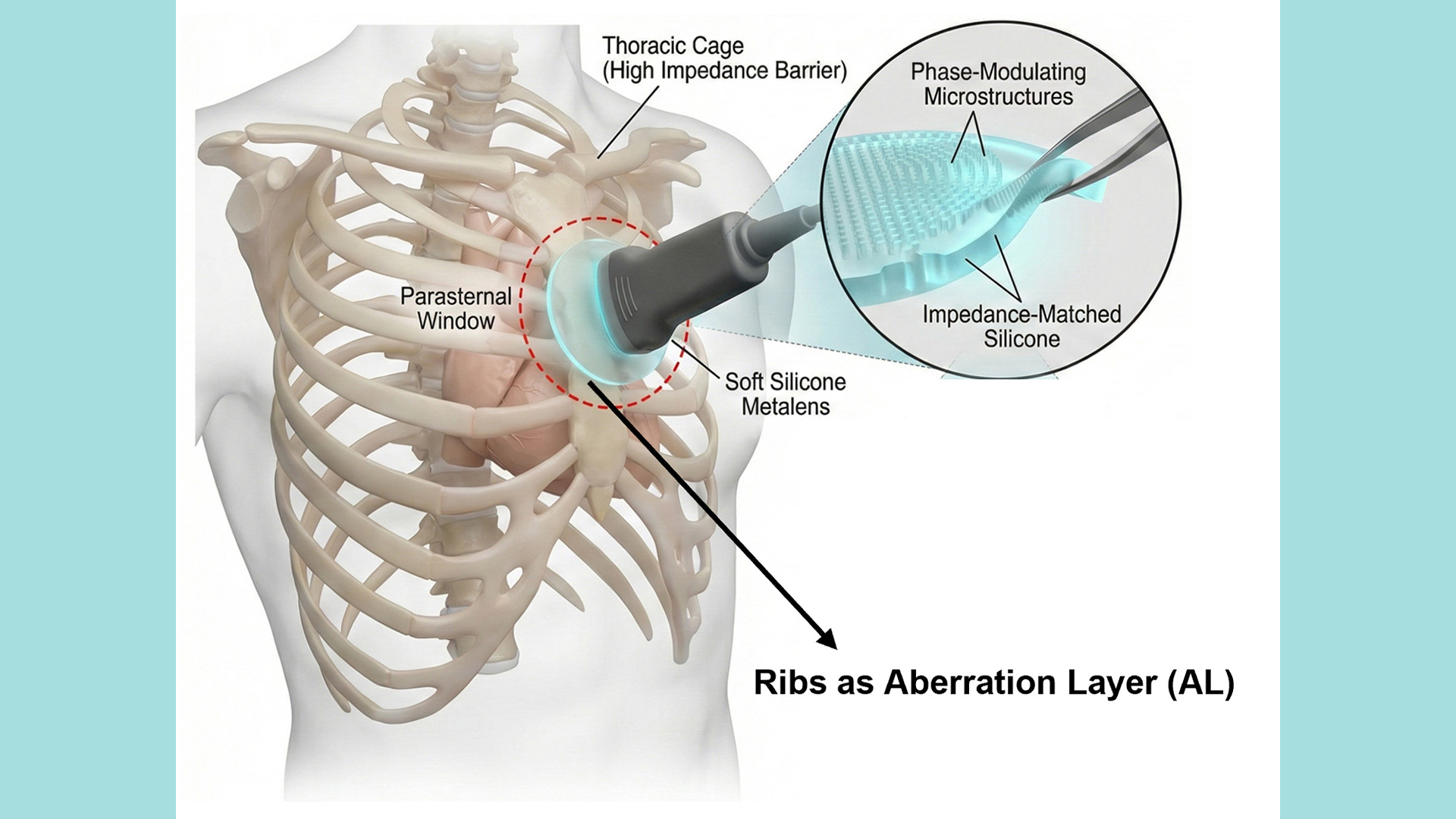
Unfortunately, traditional ultrasound examinations of the heart and lungs are often obstructed by the high impedance of the rib bones in their patients, which causes the reflection and scattering of sound waves, creating blind spots that hinder accurate and timely diagnoses. So doctors are required to scan through narrow intercostal spaces to avoid the ribs. This approach not only restricts the angles available for imaging but also complicates the acquisition of comprehensive images of the cardiac structure.
To achieve more accurate diagnoses, patients may need to be referred for X-rays or computed tomography (CT) scans. However, these methods involve radiation exposure, which is not ideal for pregnant women and patients requiring long-term monitoring. Furthermore, implementing these examinations in emergency situations can be challenging, potentially delaying critical treatment.
What is needed is a way to see past that maze of obstructions quickly and non-invasively.
Enter AI and engineering for a world-leading innovative solution named SonoMeta – an AI-powered Metalens developed by Professor Nicholas Xuanlai Fang and his research team at HKU’s Department of Mechanical Engineering that can overcome these physical hurdles and visualise beneath rib barriers.
Professor Fang and his team developed SonoMeta using cutting-edge ‘metamaterial’ technology, which employs micro-structured materials to precisely control the ultrasound waves, enabling effective penetration of rib structures while significantly reducing reflection and scattering. As a result, doctors can clearly visualise cardiac valve structures located up to approximately 10cm behind the rib cage, achieving notable improvements in imaging depth, clarity, and diagnostic accuracy compared to traditional methods.
The clinical application potential of this technology is substantial, particularly in time-sensitive environments such as emergency rooms, intensive care units (ICUs), and ambulances. Physicians will no longer be restricted by intercostal angles; they will be able to obtain complete, real-time, high-resolution images of the heart directly through the ribcage, facilitating quicker and more accurate diagnoses of critical conditions like heart failure, myocardial hypertrophy, valvular diseases, and pulmonary edema.
Moreover, SonoMeta introduces breakthroughs in the screening of pulmonary diseases. Traditional ultrasound has struggled to penetrate air-filled lung tissues and rib bones, making early detection of lung nodules or abnormalities difficult. This new Metalens allows ultrasound waves to penetrate the thoracic cavity clearly, enabling earlier identification of lung issues. In certain circumstances, it may also replace some radiation-exposing chest CT scans, significantly reducing patients’ risk of radiation exposure.
The HKU research team has successfully completed principle validation, simulation modelling, and preclinical experiments, turning this technology into the first commercialised ultrasound Metalens product globally. The team plans to refine the technology and develop a portable accessory that can be mounted on existing ultrasound probes. This advancement will enable healthcare professionals to conduct rapid, radiation-free thoracic scans anywhere, facilitating early screening for cardiovascular and cerebrovascular diseases, while expanding applications in bedside diagnostics and telemedicine.
“Our goal is to leverage innovative engineering to address significant clinical challenges,” said Professor Fang. “SonoMeta not only enhances image quality but, more importantly, makes diagnoses more precise, safe, and timely – truly embodying a patient-centred approach to healthcare.”
This groundbreaking technology is set not only to enhance diagnostic precision and efficiency but also to reduce overall medical risks and costs, opening a new chapter in the field of medical imaging worldwide.